
Once the organ extractions were freeze dried, Dr. Prior instructed us in how to the NMR spectrometer to acquire proton NMR spectra of metabolites with the tissues we had extracted. NMR spectroscopy allows us to study metabolic processes using the technique known as metabolomics, which is the “systematic study of the unique chemical fingerprints that specific cellular processes leave behind”. The NMR spectra obtained allowed us to see metabolites within the tissue extracts, however a single metabolite can give rise to several different signals, from different parts of the individual molecules, such that a methyl group (–CH3) and a methoxy (ROCH3) group, potentially causing problems with identification.
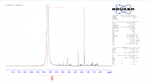
Kidney sample spectra at 16 NS
In order to prepare the samples for the NMR we decided to dissolve them in 500μl of Deuterium oxide (D2O), as this helped solubilise our extracts without introducing a large water peak, that would be visible in the spectra.
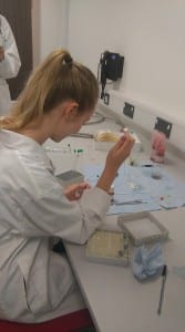
Corinne preparing the samples
The first sample to be run was the kidney sample. The acquisition parameters we used allowed us to observe signals from protons, providing a one dimensional spectra. It was found that this was very insensitive as the number of repeat scans (NS) was insufficient due to the sample not being concentrated enough. The high peak around 5.5 and 4.5 also meant that water was present with the sample.
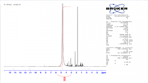
Kidney sample spectra at 512 NS
The NMR experiment was repeated; however, this time the number of scans was increased from 16 to 512. This produced a spectrum with a better signal to noise ratio, which could be analysed more easily.
Once the kidney sample scan was completed, NMR proton spectra were obtained from the other two organ extracts.
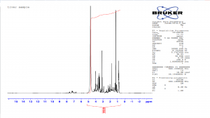
Liver sample Proton (¹H) NMR spectrum obtained with 512 scans
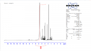
Heart sample Proton (¹H) NMR spectrum obtained with 512 scans
The liver sample provided the best (¹H) proton NMR spectrum, we believe this was because the organ was more easily broken down by the homogenisation process with the perchloric acid.
Finally, the analysis of all three (¹H) proton NMR spectra proved very interesting. There were certain similarities that could be seen in all three organs, which could be made more apparent if all the extraction were as of similar purity.
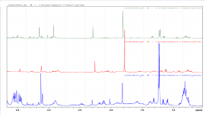
All three organ spectra compared