Category Archives: NMR

Organ Extraction Spectra

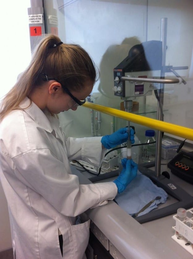
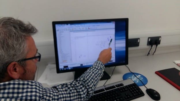
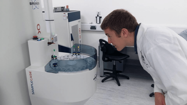
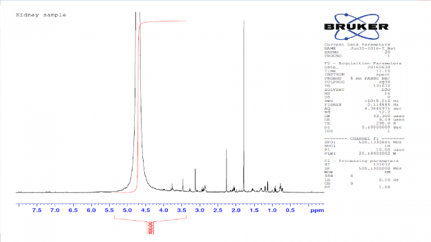
Once the organ extractions were freeze dried, Dr. Prior instructed us in how to the NMR spectrometer to acquire proton NMR spectra of metabolites with the tissues we had extracted. NMR spectroscopy allows us to study metabolic processes using the technique known as metabolomics, which is the “systematic study of the unique chemical fingerprints that specific cellular processes leave behind”. The NMR spectra obtained allowed us to see metabolites within the tissue extracts, however a single metabolite can give rise to several different signals, from different parts of the individual molecules, such that a methyl group (–CH3) and a methoxy (ROCH3) group, potentially causing problems with identification. Continue reading

Understanding Proton (¹H) NMR Spectra

Dr. Prior firstly described the mechanisms and principles behind NMR spectroscopy; in that a very strong magnetic field, maintained by a large current (60 Amps) flowing around a circular magnet (supercooled by liquid nitrogen and liquid helium “jackets”), affects the polarity of nucleus (also known as protons) within the sample. This allows biological and chemical samples with different compositions to be identified and distinguished from each other. This is because nuclear spin gives them the properties of a magnet. However, he also explained that it is highly insensitive because only one in a million protons will align with the magnetic field generated by the NMR, rather than against it.

Continue reading

Sample Preparation

As undergraduate students, it is very rare that we are afforded the opportunity to work with equipment such as the Nuclear Magnetic Resonance (NMR). Dr. Timothy Bates, our supervisor for this project, kindly organised for our group to undertake some valuable training with Dr. Stephen Prior, who is an Instrumental Scientist, at the University of Lincoln.

Continue reading