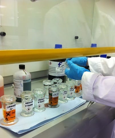
To see if the cannabinoid drugs we used had induced apoptosis (programmed cell death) in the HL-60 cells, we needed to stain them to observe their structure. When reading previous journals, it appeared that most scientists used the Giemsa stain; however, we wanted to see if two other stains would also produce useful data.
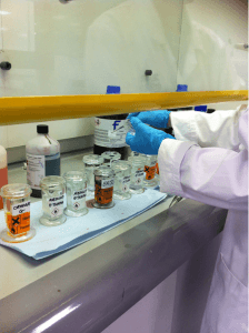
Lee-Anne staining the slides with the HL60 cells.
First we used methylene blue stain in order to try and observe the HL60 cells’ nuclei. This proved unsuccessful, as despite leaving the cell samples in the methylene blue for 8 minutes, the cellular structures did not appear to take up the stain and very little could be seen under the light microscope.
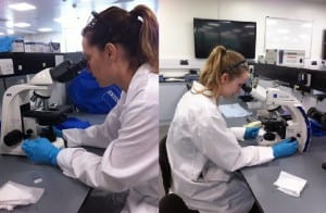
Lee-Anne and Corinne observing the stained HL60 cells through the Zeiss Primo Star Laboratory Microscope.
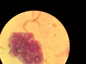
The next stain we used was the Papanicolaou stain, as this contains multiple dyes to differentially stain various components of the cell. Haematoxylin stains the nucleus, orange G stains keratin and EA 50 the cytoplasm. The protocol we followed was that of a previous university practical used with cells obtained from the human cheek. The Papanicolaou stain gave a better result than the methylene blue, but the cells were still very light under the microscope, making it harder to observe apoptosis. We then extended the time that the HL-60 cells were exposed to the stain to see if this would give better results, however it did not.
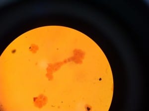
HL60 cells stained with Papanicolaou stain.
Finally we used the Giemsa stain, which is specific to the phosphate group of DNA, attaching itself to the regions where high amounts of adenine and thymine are bonded. This stain gave the best results, as we could clearly observe the nucleus and cell membrane. This allowed us to see if apoptosis had occurred. As suggested in the journals, 10 minutes was ample time for the stain to take effect.
HL60 cells stained with Giemsa stain, which provided a much clearer image.
Once staining had taken place we were able to use the Nikon Eclipse E-800 upright microscope to obtain clearer images of our cells. This was very exciting as we could see if the cannabinoids we used had induced apoptosis, which in some cases it had.
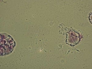
HL60 cells having undergone apoptosis. Giemsa stained.